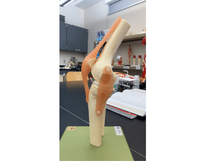
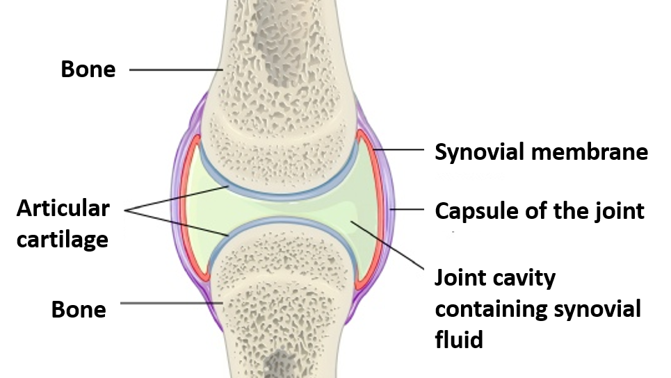
Ever wondered what makes your knee so complex and crucial to your movement? This amazing joint, connecting your thigh to your lower leg, is a masterpiece of engineering, allowing us to walk, run, jump, and even dance. But navigating the intricate network of bones, ligaments, tendons, and cartilage can be daunting. So, let’s delve into the fascinating world of the knee joint and learn how to label its vital components.

Image: www.purposegames.com
This comprehensive guide will serve as your roadmap to unraveling the mysteries of the knee joint. We’ll explore the key structures, their interconnected functions, and the importance of understanding them. Whether you’re a healthcare professional, a fitness enthusiast, or simply curious about the mechanics of your own body, this journey will equip you with valuable knowledge about this remarkable joint.
The Foundation: Bones and Their Connections
The Three Main Bones
The foundation of the knee joint rests on three crucial bones: the femur (thigh bone), the tibia (shin bone), and the patella (kneecap). The femur, the longest and strongest bone in the body, serves as the upper link, while the tibia acts as the lower link for the knee joint. The patella, a small, triangular bone that sits in front of the knee, protects the joint and acts as a lever to amplify the force during extension.
The Patellofemoral Joint
The articulation between the patella and the femur forms the patellofemoral joint. This joint plays a vital role in knee extension by redirecting the forces generated by the quadriceps muscle. It allows the kneecap to glide smoothly within the groove of the femur, preventing friction and ensuring efficient movement.

Image: denisgeeks.weebly.com
The Tibiofemoral Joint
The main articulation of the knee, the tibiofemoral joint, is where the femur meets the tibia. This joint is responsible for flexion and extension movements, enabling the bending and straightening of the knee. It is also responsible for some rotational movements, though these are limited compared to other joints in the body.
The Crucial Connectors: Ligaments
The knee joint is held together and stabilized by a network of strong, fibrous ligaments that restrict excessive movement and prevent injury. Each ligament serves a unique role in maintaining joint stability. Let’s examine some of the key ligaments:
The Anterior Cruciate Ligament (ACL)
The ACL is a vital ligament that connects the front of the tibia to the back of the femur. This ligament prevents the tibia from sliding forward relative to the femur, a crucial function during activities involving pivoting, jumping, and landing. The ACL is often injured during sports or falls due to sudden twisting or impact forces.
The Posterior Cruciate Ligament (PCL)
The PCL is positioned on the back of the knee, connecting the back of the tibia to the front of the femur. Its primary role is to prevent the tibia from sliding backward relative to the femur. The PCL is less prone to injury than the ACL but can be affected by forceful hyperextension or direct impact to the knee.
The Medial Collateral Ligament (MCL)
The MCL is located on the inner side of the knee, running along the edge of the tibia and femur. It provides medial stability, preventing the knee from bending inward. The MCL is commonly injured during direct blows to the outer side of the knee, such as during contact sports.
The Lateral Collateral Ligament (LCL)
The LCL is situated on the outer side of the knee, connecting the head of the fibula to the femur. Similar to the MCL, it acts as a restraint against lateral forces, preventing the knee from bending outward. The LCL is more prone to injury due to direct blows to the inner side of the knee or severe twisting movements.
The Movers and Shakers: Muscles and Tendons
The knee’s movement is orchestrated by a complex interplay of muscles and tendons. These structures generate force and transmit it to the bones, allowing for a full range of motion. Let’s explore some of the major players:
Quadriceps Muscle
The quadriceps muscle group, located on the front of the thigh, consists of four muscles: the rectus femoris, vastus lateralis, vastus medialis, and vastus intermedius. This group is responsible for extending the knee and is essential for activities like walking, running, and jumping. The quadriceps muscle connects to the patella via the patellar tendon, further amplifying its force during knee extension.
Hamstring Muscles
The hamstring muscle group, positioned on the back of the thigh, consists of three muscles: the biceps femoris, semitendinosus, and semimembranosus. These muscles work in opposition to the quadriceps, flexing the knee and enabling back-bending movements.
Popliteus Muscle
The popliteus muscle, located on the back of the knee, plays a crucial role in initiating knee flexion and externally rotating the tibia. It also supports stability by preventing the femur from sliding off the tibia during flexion.
Other Important Muscles
Other muscles contribute to knee function, including the gastrocnemius (calf muscle) and the soleus. These muscles, located in the lower leg, support knee flexion and help with plantarflexion (pointing the toes downward).
The Protective Layer: Cartilage
The articular cartilage, a smooth, slippery layer of tissue that covers the ends of the femur, tibia, and patella, plays a crucial role in protecting the knee joint. This cartilage cushions the bones, reduces friction during movement, and ensures smooth gliding between the joint surfaces. It absorbs shock and prevents wear and tear on the bones, keeping the knee functioning efficiently.
Types of Articular Cartilage
The knee joint houses two distinct types of articular cartilage: hyaline cartilage and fibrocartilage. Hyaline cartilage, found on the surfaces of the femur, tibia, and patella, provides smooth gliding and shock absorption. Fibrocartilage, located in the menisci, offers additional support and stability to the joint.
The Menisci: Cushions and Stabilizers
The menisci, C-shaped pieces of fibrocartilage, reside within the tibiofemoral joint. These structures play a crucial role in distributing weight, absorbing shock, and providing stability to the knee. Each knee has two menisci: the medial meniscus on the inner side and the lateral meniscus on the outer side.
Crucial Functions of the Menisci
The menisci perform numerous vital functions:
- Shock Absorption: They act as cushions to distribute the body’s weight, reducing stress on the joint surfaces during activities like walking, running, and jumping.
- Joint Stability: They help stabilize the knee, preventing excessive movement and potential injuries.
- Lubrication: They facilitate smooth gliding between the femur and tibia during movement.
- Joint Congruence: They help improve the fit between the femur and tibia, enhancing the stability and efficiency of the joint.
Meniscus Injuries
The menisci are susceptible to injuries due to their role in absorbing shock and providing stability. Tears or degeneration of the menisci can occur during sports, falls, or even with age. Meniscus injuries can cause pain, swelling, and a range of other symptoms, depending on the severity and location of the tear.
A Glimpse into the Knee’s Complexity
The knee joint, a remarkable masterpiece of engineering, is a testament to the intricate design of the human body. By understanding each component and its function, we gain a deeper appreciation for the complexity and elegance of this crucial joint. From the sturdy bones to the delicate ligaments, powerful muscles, and resilient cartilage, every structure plays a critical role in enabling movement, providing stability, and protecting the joint from injury.
Importance of Knee Joint Labeling
The ability to accurately label the components of the knee joint is essential for a wide range of individuals, including:
- Healthcare Professionals: Doctors, nurses, physical therapists, and athletic trainers rely on precise anatomical knowledge to diagnose and treat knee injuries, perform surgery, and design rehabilitation plans.
- Patients: Understanding the anatomy helps patients comprehend their condition, participate actively in their treatment, and make informed decisions about their healthcare.
- Fitness Enthusiasts: Athletes, fitness trainers, and exercise instructors benefit from a thorough understanding of the knee joint to develop safe and effective training programs, prevent injuries, and optimize performance.
- General Public: Knowing the basic components of the knee helps us make healthier lifestyle choices, recognize potential warning signs of injuries, and take preventive measures to preserve joint health over time.
Knee Joint Labeling
Conclusion: Navigating the Knee Joint
This comprehensive labeling guide has provided a detailed look at the intricate anatomy of the knee joint. By exploring each component, its function, and its vital role in joint stability and movement, we gained a deeper understanding of this remarkable structure. The knee joint, with its complex interplay of bones, ligaments, muscles, and cartilage, is a testament to the efficiency and brilliance of human anatomy. By appreciating this complexity, we can better understand the potential for injury and learn to protect this precious joint through proper care, exercise, and preventative measures.


